Scientists use intracranial EEG technology to reveal human low-frequency θ oscillations carrying grid cell information
October 24, 2018 Source: Chinese Academy of Sciences
Window._bd_share_config={ "common":{ "bdSnsKey":{ },"bdText":"","bdMini":"2","bdMiniList":false,"bdPic":"","bdStyle":" 0","bdSize":"16"},"share":{ }};with(document)0[(getElementsByTagName('head')[0]||body).appendChild(createElement('script')) .src='http://bdimg.share.baidu.com/static/api/js/share.js?v=89860593.js?cdnversion='+~(-new Date()/36e5)];When the animal is free to explore the external environment, the Grid cell in the Entorhinal cortex of the brain participates in it and presents a unique hexagonal discharge pattern in space (Fig. 1a). This discovery has attracted the interest of many neuroscientists since it was first reported in 2005. Subsequent studies have further found that grid cells play an important role in the function of the brain to characterize spatial location and complete path integration. Grid cells open the way for people to understand the cerebral cortex. In 2014, the founder of the grid cell Moser and his mentor John O'keefe (discovered location cells) shared the Nobel Prize in Physiology or Medicine for revealing the brain GPS positioning system.
The generation of neuronal action potentials is closely related to the field potential activity of local neuronal populations. Animal and human electrophysiological studies have found that the entorhinal cortex exhibits strong θ oscillations (4-8 hz) during spatial navigation. The results of computational modeling suggest that the hexagonal pattern of the grid cells is mainly formed by the θ oscillation superimposed interference of the entorhinal cortex. A large number of experimental studies by predecessors have also shown that θ oscillations can encode specific spatial information. For example, θ oscillation can encode the motion speed, boundary distance, and the like of the subject. Human fMRI studies have also confirmed the presence of signals for the Gird cell-like representation in the entorhinal cortex. However, what is the neuroelectrophysiological basis of this type of grid cell representation in the human brain is still unresolved. Answering this question will help to further understand the formation of grid cells and how the brain transmits grid signals.
Researchers from the Institute of Psychology of the Chinese Academy of Sciences, Beijing Normal University, and Ruhr University in Bochum, Germany, recruited nine hospitalized patients with epilepsy (from the Yuquan Hospital affiliated to Tsinghua University, the First Affiliated Hospital of the People's Liberation Army General Hospital, and the University of Freiburg, Germany). Hospital), they need surgery for invalid medical treatment. The preoperative doctor will implant multiple deep SEEG electrodes in the patient's brain to accurately assess the location of the epileptic focus. The position of the electrode implantation is determined entirely according to the purpose of the treatment, and has nothing to do with the study. During the experiment, the subject needed to complete a virtual reality task related to object-position-related memory (Fig. 1c). At the same time, the researchers directly recorded the field potential signal of the cerebral cortex through the intracranial electrode, and then used parametric linear regression. The method calculates the relationship between the virtual moving direction and the signal strength of different frequencies of the entorhinal cortex. Previous studies have suggested that when the grid cells successfully encode the spatial position, the neuron firing will form six main axes with a 60-degree interval (Fig. 1a black line): when the subject moves along the main axis, the grid cells discharge more. Intense, while moving in the non-spindle direction, the grid cells discharge is relatively weak. The study assumes that the amplitude of the neural oscillation signal is modulated by the direction of motion: there is a six-cycle rotational symmetry between the amplitude and the direction of motion (Fig. 1b).
The researchers first studied whether the θ oscillation of the entorhinal cortex (the black point in Figure 2a is the electrode contact located in the entorhinal cortex) carries the grid cell signal. As shown in Fig. 2b, after the amplitude of the θ oscillation is divided into 12 parts according to the direction of motion of the subject, a significant six-period mode is exhibited, and the height between the high and low is exactly 60 degrees, which proves the θ oscillation of the human entorhinal cortex. Carry a grid cell signal. In order to further study whether the six-cycle mode exists only in the θ band, the researchers extract the δ band (2-4hz), the α band (8-12hz), the β band (12-30hz), the low frequency γ band (30-80hz) and the high. Frequency gamma band (80-150hz) signal and analyzed. However, the researchers found that the amplitudes of these bands did not appear in the six-cycle mode, and the modulation intensity was not significant (Fig. 2c). These results show that only θ oscillations can carry a grid signal. Is the grid signal unique to the entorhinal cortex? The researchers used the same method to study the hippocampus and amygdala adjacent to the entorhinal cortex, and found no six-cycle modulation pattern (Fig. 2d).
Animal studies have found that as the mouse becomes more familiar with the spatial environment, the hexagonal pattern of grid cells is better characterized. The closer the grid cells are to the boundary, the more regular the hexagonal pattern, indicating that the boundary has an anchoring effect on the grid. Does human grid cells have similar properties? The researchers further explored these two issues. The study divided the experimental duration into six equal sessions, and calculated the strength of the six-cycle modulated signal from session 1/2 to session 3/4 and session 3/4 to session 5/6. The researchers found that only the six-cycle modulation signal from session 3/4 to session 5/6 was significantly larger than zero, indicating that the grid representation of the subjects tends to be stable in the later stages of the experiment (Fig. 2e), indicating that the grid cells need to be familiar with the subjects. Only after the environment can there be better coding space. In addition, the researchers divided the space into three parts of equal radius according to the range of activities of the subjects: Border, Middle and Inner, respectively, and calculated the intensity of the six-cycle modulated signals from Border to Middle and Middle to Inner. The study found that only the border-to-Middle six-cycle modulation signal is significantly larger than zero, indicating that the mesh representation of the subject is more stable in the boundary region than in the central region (Fig. 2f), revealing that the virtual boundary also has an anchoring effect on the mesh mode.
Based on the unique discharge mode of grid cells, the electrophysiological signal is modulated by the 6-cycle rotational symmetry of the motion direction. It is shown for the first time that θ oscillation can also encode grid signals, and the first time in human subjects, grid representation is found in time. Progressive stability above, the boundary area in space is more stable than the central area. At the same time, the study also confirmed for the first time the existence of neural oscillations based on fMRI-based characterization of cell-like cells.
The research was mainly done by researchers from the Institute of Psychology, Beijing Normal University and Ruhr University in Bochum, Germany. PhD student Chen Dong and Dr. Lukas Kunz are the first authors, and Professor Nikolai Axmacher and researcher Wang Liang (as lead contact) are co-directed authors. The research was funded by the National Youth Science Foundation Outstanding Youth Fund (81422024), the Beijing Municipal Science and Technology Commission Brain Cognition and Brain Medicine (Z171100000117014) and the Chinese Academy of Sciences Mental Health Key Laboratory Project (KLMH2018ZK02). The paper was published online on October 11th in the Current Biology magazine of Cell.
Paper information: Chen D, Kunz L, Wang W, Zhang H, Wang W, Schulze-Bonhage A, Reinacher PC, Zhou W, Liang S, Axmacher N, Wang L. Hexadirectional modulation of theta power in human entorhinal cortex Current Biology 2018. DOI: https://doi.org/10.1016/j.cub.2018.08.029
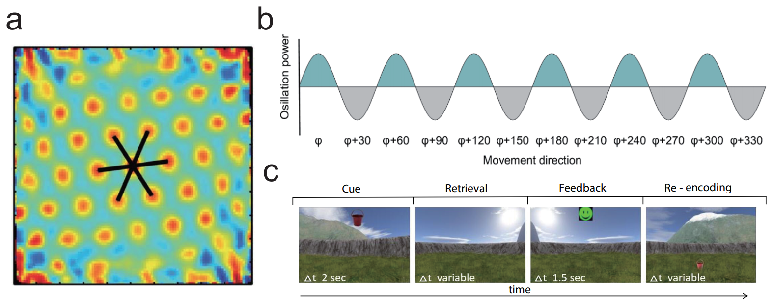
figure 1

figure 2
Canned Mandarin Oranges are processed with Satsuma variety orange, which is an unique variety in China, famous for it's thin peel, seedless and good flavor. It's a good source of Vitamin C.
Every piece of fresh mandarin orange is selected and pesticides & heavy metal residual tested. Our Canned Mandarin Oranges contain no Carbendazim.
During the period of planting, we provide the pesticides for farmers, and professional staffs from the company will guide farmers on how to use and spray pesticides scientifically. After usage, the rest pesticides and pesticides containers will be stored and disposed correctly.

With processed and shelf-stable canned mandarin orange, you can enjoy fresh flavors of fresh mandarin orange at anytime and anywhere.
We have different pack sizes, package material and packing medium for your choice.
Canned Orange,Canned Mandarin Orange,Mandarin Oranges In Syrup,Mandarin Oranges In Light Syrup
ZHEJIANG FOMDAS FOODS CO., LTD. , https://www.fomdasfoods.com